
Requires;
1 piece of long Y TAPE
1 piece of short Y TAPE
Self-taping is available
Step 1.

Step 2.

Step 3.
No stretch is required during application.
Kinesio tape, Kinesio tex tape, Kinesiology tape, Sports tape, KT Tape, Rock tape, Athletic tape, Medical tape, Muscle tape, Trainers tape, Physical therapy tape, Health Care Product, Pharmacy Stocks, Balance Tape, Bandage, Elastic Tape, Rehabilitation, Sports Goods, Fitness Product, Orthopedic
Description
The ankle joint is one of the most commonly injured joints in the body with the lateral ankle sprain being the most frequent type of sprain. The mechanism of injury is when the foot is in an inverted position combined with plantar flexion which usually does damage to the lateral complex of the ankle.1,2
Anatomy
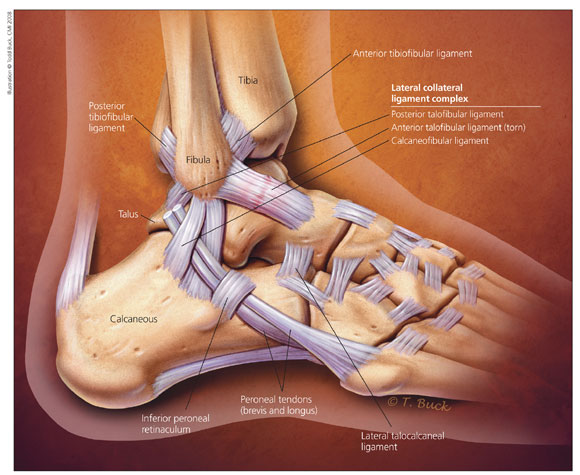
The ankle joint is the most congruent in the body and it is due to the bony articulations, ligaments, capsule, and tendons. The lateral complex of the ankle is made up of three separate ligaments. The anterior talo-fibular ligament (ATFL) is the weakest out of the three ligaments and the most common to be injured. Its role is to limit plantar flexion and inversion of the ankle which is why it is so susceptible to injury because of the common mechanism of injury. The other two ligaments are the calcaneo-fibular ligament (CFL) and the posterior talo-fibular ligament (PTFL). The posterior talo-fibular ligament is the strongest of the three ligaments and is rarely injured.3-5
Classifications
Lateral ankle sprain can be categorized into three different grades:
Grade I (mild): There is a mild stretch to the ATFL, which is usually the only ligament involved in a grade I. Some ligament fibers may be torn but there is no to little limp, mild swelling, and minimal functional loss.
Grade II (moderate): There is a complete tear of the ATFL and a partial tear to the CFL. There is an inability to toe raise and a limp with walking is developed. Range of motion is restricted with swelling and tenderness.
Grade III (severe): There is a complete tear of the ATFL and CFL and a partial tear of the PTFL. Initially, there is complete loss of range of motion and an inability to weight bear.3,5,6
Incidence and Prevalence
Ankle sprain is the most common sports-related injury due to all the force it has to withstand. It is typical to see an ankle sprain on the dominant leg, and it is regularly the lateral ligament complex that is injured. It is likely to see these injuries with sports that require running, balance, cutting, and stop-and-go movements such as basketball, soccer, and football.1,3-7
Clinical Presentations
After an ankle injury, the athlete will usually describe the mechanism of injury as "rolling" his or her ankle. The picture below demonstrates the common mechanism of injury for a lateral ankle sprain.

Depending on the grade, the athlete is unlikely to continue playing their sport. Some of the clinical symptoms one will see are the following:3,8
• Swelling
• Pain
• Bruising
• Sensory nerve damage
• Difficultly bearing weight
• Instability
Diagnostic Tests
A radiograph is not used to diagnose an ankle sprain, but it is used to rule out fractures. The Ottawa Ankle Rules are used as a predictor for patients with an ankle sprain that should have an x-ray. The three rules are the following:3,4,6,9,10
• Bony tenderness at the base of the 5th metatarsal
• Inability to bear weight immediately after the injury and for four steps
• Bony tenderness at the tip or posterior edge of the malleolus
Stress radiographs can be used to diagnose between a single and multiple ligament injury. However, it is not generally used because the information is not clinically useful and there is a lack of universally accepted criteria.2,3,6 A magnetic resonance imaging is accurate and reliable to determine ligament damage, but this is used more for chronic ankle instability and usually an expensive approach.3,11
Evaluation/Special Orthopedic Tests
The two stress tests that are used to assess injury to the lateral ligaments are the anterior drawer test, which assess the ATF, and talar tilt test, which assesses both ATF and CFL. It is important to compare bilaterally to see the difference of laxity between both ankles. Using these laxity tests may not be accurate initially with pain and swelling. The test should be repeated again after pain and swelling has gone down. To perform the anterior drawer test, the examiner stabilizes the tibia and fibula, and draws the talus forward in the ankle mortise. For the talar tilt test, the examiner is still stabilizing the tibia and fibula, but the ankle joint is being pushed into inversion, stressing the lateral complex. If the patient presents with excessive laxity and/or pain, the test is positive for a ligamentous tear. The following are videos on how to perform these laxity tests.
Conservative Treatment
It is normal to do conservative treatment with patients with lateral ankle sprains and the prognosis is usually a good outcome. The grade of injury usual dictates how conservative treatment will be performed. Grade III sprains are also treated conservatively but on the rare occasions, it could also be treated surgically.3,5,9,10 Regardless of grade of ankle sprain, initially the ankle will be treated with the PRICE principle (protection, rest, ice, compression, and elevation). During this short period of immobilization, the following equipment can help achieve the initial goal of reducing pain and swelling: elastic or inelastic tape or bandage, semi-rigid support, lace-up ankle braces, and/or crutches. Other modalities that can be used initially are cryotherapy, non-steroidal anti-inflammatory drugs, ultrasound and interferential.3,5,9,10,12,13 Click on the physical therapy management of lateral ankle sprain link for more information on the modalities that is generally used in physical therapy practice.
For grade II and III ankle sprains, once the patient gets into the sub-acute stage of rehabilitation, the treatment can be directed towards increasing pain-free motion and general ankle muscle strengthening. The last phase of treatment involves proprioceptive exercises, which is a key component of not re-injuring the ankle. Proprioceptive training with a wobble board, mini tramps, or rocker boards should start after 3 to 4 weeks of rehabilitation. External supports should be used to provide proprioceptive feedback in order to improve balance and neuromuscular control of the ankle.3,5,6,9,10,12
Click on the link below to read about current evidence-based practice of a lateral ankle sprain.
Physical Therapy Management of Lateral Ankle Sprain
Surgery and Postoperative Treatment
Surgery is not recommended for acute ankle sprain and is mainly used to manage chronic lateral ankle instability. Another indication for surgery is if conservative methods have failed. If surgery is needed, the three common types of surgery are anatomic repair, nonanatomic reconstruction, and anatomic reconstruction. Postoperative protocol for operative treatment has the patient in a splint for the first two weeks after surgery. Then ankle is neutral and foot everted. Gentle stretching begins but with no passive inversion stretching for six weeks. After this point, the protocol that is used for conservative treatment is the same protocol that will be used for the rest of the operative treatment. Ankle bracing is required full-time for the initial three months and is always recommended for high risk activities.3,5-7
Additional Web-based Sources
http://orthopedics.about.com/cs/sprainsstrains/a/anklesprain.htm
http://www.wheelessonline.com/ortho/ankle_sprain
http://www.mckinley.illinois.edu/handouts/anklesprain/anklesprain.html
http://www.healthychildren.org/English/health-issues/injuries-emergencies/sports-injuries/pages/Lateral-Ankle-Sprain-and-Rehabilitation.aspx?nfstatus=401&nftoken=00000000-0000-0000-0000-000000000000&nfstatusdescription=ERROR%3a+No+local+token
http://www.upmc.com/Services/sportsmedicine/injuries/leg/Pages/ankle-sprain.aspx
References
http://morphopedics.wikidot.com/lateral-ankle-sprain
1. Morrison KE, Kaminski TW. Foot characteristics in association with inversion ankle injury. J Athl Train. 2007;42(1):135-142.
2. Bennett WF. Lateral ankle sprains. part I: Anatomy, biomechanics, diagnosis, and natural history. Orthop Rev. 1994;23(5):381-387. http://search.ebscohost.com/login.aspx?direct=true&db=cmedm&AN=8041572&site=ehost-live.
3. Chan KW, Ding BC, Mroczek KJ. Acute and chronic lateral ankle instability in the athlete. Bull NYU Hosp Jt Dis. 2011;69(1):17-26. http://search.ebscohost.com/login.aspx?direct=true&db=cmedm&AN=21332435&site=ehost-live.
4. Lynam L. Assessment of acute foot and ankle sprains. Emerg Nurse. 2006;14(4):24-33. http://search.ebscohost.com/login.aspx?direct=true&db=cmedm&AN=16878848&site=ehost-live.
5. Pollard H, Sim P, McHardy A. Lateral ankle injury. literature review and report of two cases. Australas Chiropr Osteopathy. 2002;10(1):21-30. http://search.ebscohost.com/login.aspx?direct=true&db=cmedm&AN=17987171&site=ehost-live.
6. Ferran NA, Oliva F, Maffulli N. Ankle instability. Sports Med Arthrosc. 2009;17(2):139-145. http://search.ebscohost.com/login.aspx?direct=true&db=cmedm&AN=19440141&site=ehost-live.
7. Cohen RS, Balcom TA. Current treatment options for ankle injuries: Lateral ankle sprain, achilles tendonitis, and achilles rupture. Curr Sports Med Rep. 2003;2(5):251-254.
8. Cardle I. The treatment and prevention of common ankle injuries. Peak Performance. 2009. http://files.pponline.co.uk/102_downloads/the_treatment_and_prevention_of_common_ankle_injuries.pdf
9. Lynch SA, Renström ,P.A. Treatment of acute lateral ankle ligament rupture in the athlete. conservative versus surgical treatment. Sports Med. 1999;27(1):61-71. http://search.ebscohost.com/login.aspx?direct=true&db=cmedm&AN=10028133&site=ehost-live.
10. Puffer JC. The sprained ankle. Clin Cornerstone. 2001;3(5):38-49. http://search.ebscohost.com/login.aspx?direct=true&db=cmedm&AN=11464730&site=ehost-live.
11. Kirk T, Saha S, Bowman LS. A new ankle laxity tester and its use in the measurement of the effectiveness of taping. Med Eng Phys. 2000;22(10):723-731.
12. Slimmon D, Brukner P. Sports ankle injuries - assessment and management. Aust Fam Physician. 2010;39(1-2):18-22. http://search.ebscohost.com/login.aspx?direct=true&db=cmedm&AN=20369129&site=ehost-live.
13. Lin CC, Hiller CE, de Bie R,A. Evidence-based treatment for ankle injuries: A clinical perspective. J Man Manip Ther. 2010;18(1):22-28. http://search.ebscohost.com/login.aspx?direct=true&db=cmedm&AN=21655420&site=ehost-live.
That would extend my reflexes. Great tips!
답글삭제grout cleaning phoenix az
Thanks for sharing this information. I found it very informative as I have been researching a lot lately on practical matters such as you talk about..
답글삭제Sports Tape Australia
There's shocking news in the sports betting industry.
답글삭제It's been said that every bettor must watch this,
Watch this now or quit placing bets on sports...
Sports Cash System - Advanced Sports Betting Software
QUANTUM BINARY SIGNALS
답글삭제Professional trading signals sent to your mobile phone every day.
Follow our trades today & make up to 270% daily.